Quick Navigation:
| | | |
The Circulatory System
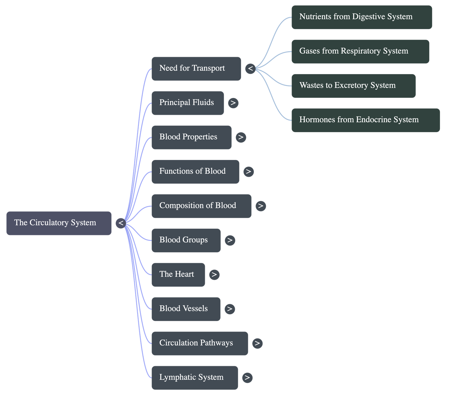
8.1 Need for Transport Inside the Body
- Every living organism requires a system to distribute essential substances and collect wastes from cells.
- Digestive system: Needs a medium to transport absorbed nutrients to every cell.
- Respiratory system: Requires a mechanism to deliver oxygen to tissues and carry carbon dioxide back to the lungs.
- Excretory system: Relies on transport to move metabolic wastes (like urea) from body parts to the kidneys.
- Endocrine system: Needs to transport hormones directly to target organs throughout the body.
8.2 Fluids in Our Body
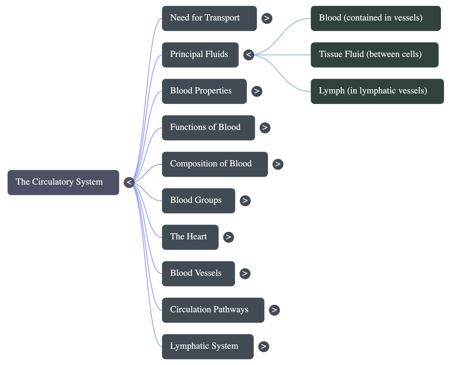
- Blood: Contained within the heart and blood vessels (closed circulatory system).
- Tissue fluid: Occupies the spaces between cells within organs.
- Lymph: Found within lymph vessels and lymphatic organs like the spleen and tonsils.
- Non-circulating fluids: Synovial fluid (in joints) and vitreous humour (in the eye).
8.3 Properties of Blood
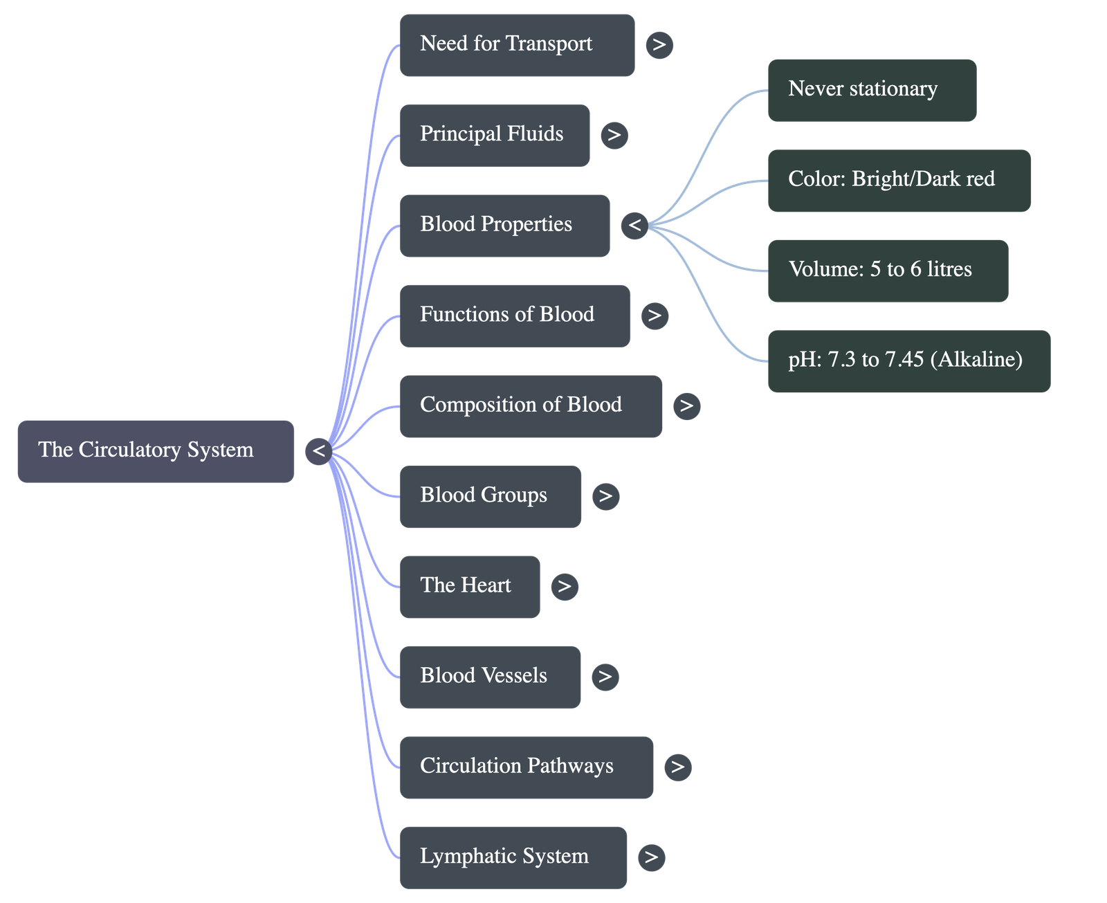
- Never stationary: It is always in continuous motion from the heart to vessels and back.
- Colour: Bright red when oxygenated (arteries) and dark red when deoxygenated (veins).
- Volume: An average adult human has about 5 to 6 litres of blood.
- Taste & pH: Saltish in taste and slightly alkaline with a pH range of 7.3 to 7.45.
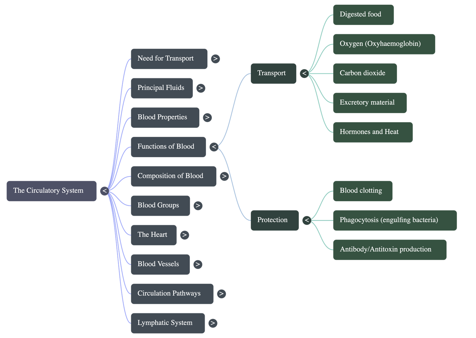
8.4 Functions of Blood
A. Transport by Blood
- Transports digested food (glucose, amino acids) to tissues.
- Transports oxygen from lungs to tissues via oxyhaemoglobin.
- Transports carbon dioxide from tissues to lungs via carbaminohaemoglobin and plasma.
- Carries excretory wastes to the liver, kidneys, or skin.
- Distributes hormones secreted by endocrine glands.
- Distributes heat to maintain uniform body temperature.
B. Protection by Blood
- Forms clots to prevent further blood loss and block entry of disease-causing germs.
- White Blood Cells (WBCs) engulf and destroy bacteria.
- Produces antibodies and antitoxins to neutralize poisons and kill germs.
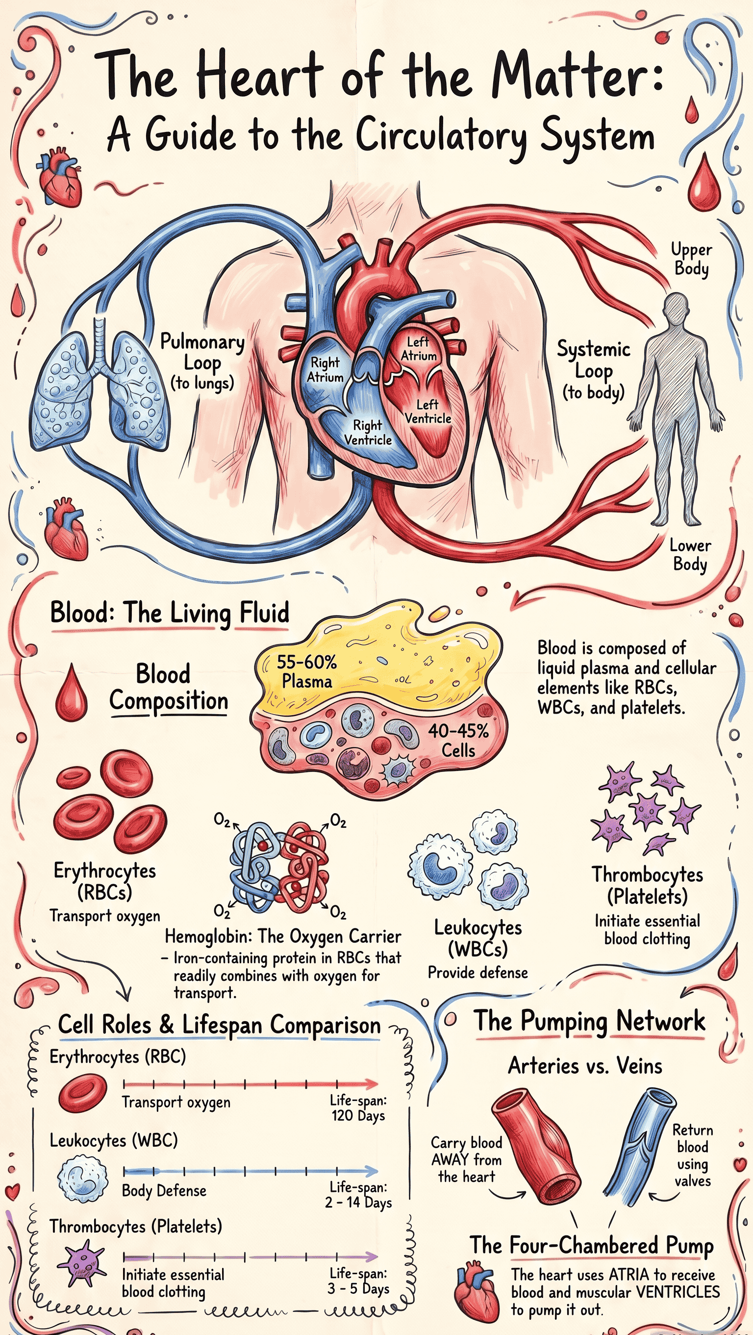
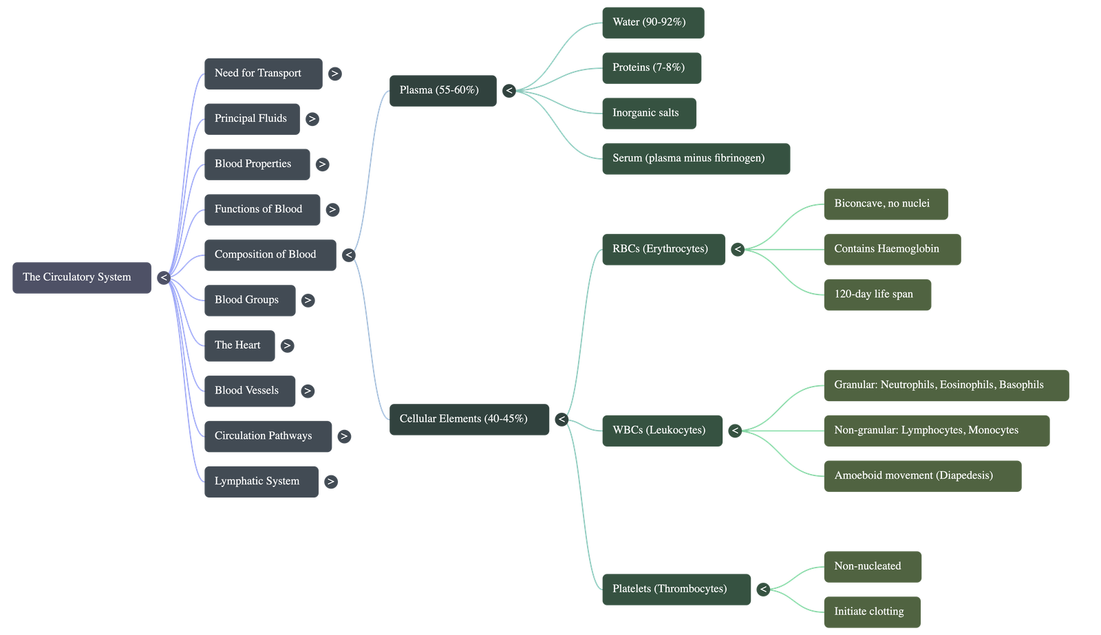
8.5 Composition of Blood
- Plasma (55-60%): The light-yellow liquid portion. Consists of 90-92% water, 7-8% proteins, and 1% inorganic salts (sodium chloride, sodium bicarbonate). Plasma minus fibrinogen is called serum.
- Cellular Elements (40-45%): Includes RBCs, WBCs, and Platelets.
Red Blood Cells (Erythrocytes)
- Minute, biconcave disc-like structures. Count: ~5 million/mm³ in adult males, ~4.5 million/mm³ in females.
- Contain haemoglobin, formed of an iron part (haemin) and protein (globin). Combines with oxygen to form unstable oxyhaemoglobin.
- Produced in bone marrow. Average lifespan is 120 days. Destroyed in the spleen, liver, and bone marrow.
- Mammalian RBC efficiency: Mature RBCs lack a nucleus (more surface area for oxygen), lack mitochondria (don't consume the oxygen they carry), and lack endoplasmic reticulum (more flexibility to squeeze through capillaries).
White Blood Cells (Leukocytes)
- Nucleated cells, lacking haemoglobin. Count: 4000-8000/mm³.
- Capable of amoeboid movement and can squeeze out of capillaries (diapedesis).
- Granular WBCs: Neutrophils (phagocytosis), Eosinophils (allergy response), Basophils (release histamine).
- Non-granular WBCs: Lymphocytes (produce antibodies) and Monocytes (ingest germs, transform into macrophages).
- Functions include phagocytosis (engulfing germs), inflammation (local heat, redness, swelling for defense), and forming antibodies (immunity).
8.6 Functions of Platelets - Clotting of Blood
- Platelets (Thrombocytes): Minute, round, non-nucleated structures. Count: 200,000 to 400,000/mm³. Lifespan is 3-5 days.
- Clotting Process:
- Injured tissues/platelets release thrombokinase.
- Thrombokinase, with calcium ions, converts inactive prothrombin into active thrombin (Vitamin K is essential for prothrombin production).
- Thrombin converts soluble plasma fibrinogen into an insoluble fibrin mesh.
- Blood cells trap in the mesh, forming a clot (thrombus). The clear liquid left is serum.
- Haemophilia: A genetic disorder where blood does not clot properly.
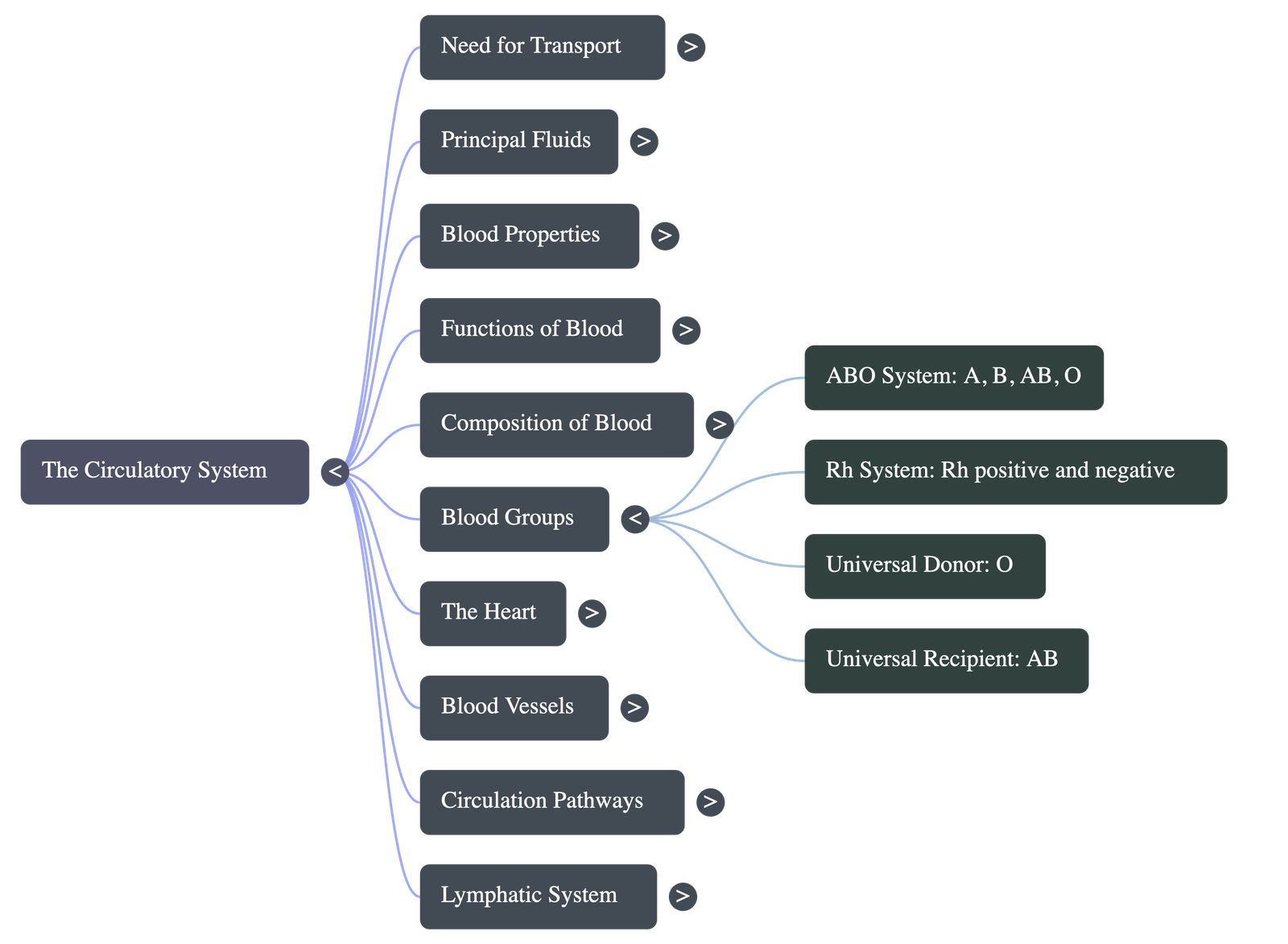
8.7 Blood Transfusion and Blood Groups
- ABO System: Blood classified into A, B, AB, and O based on antigens on RBCs and antibodies in plasma.
- Blood Group O is the universal donor; Blood Group AB is the universal recipient.
- Rh System: Presence (Rh+) or absence (Rh-) of Rh factor (Rhesus antigen) on RBCs.
- An Rh-negative mother carrying an Rh-positive foetus can develop complications (sensitization) during her second pregnancy, sometimes leading to foetal death.
8.8 Blood Circulatory System
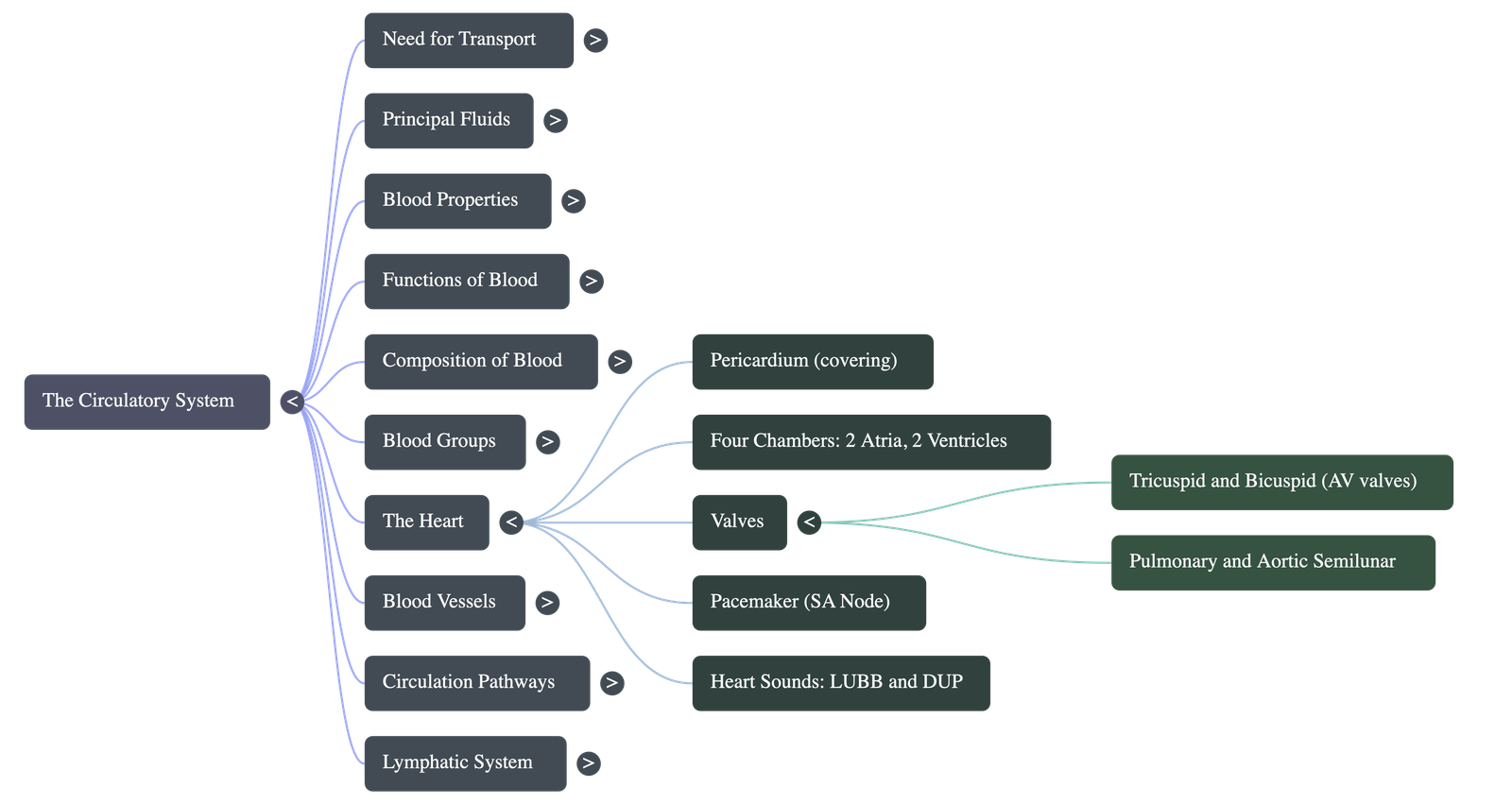
8.8.1 The Heart
- Centrally located between lungs, tilted left. Protected by double-walled pericardium with lubricating pericardial fluid.
- Chambers: Two upper atria (thin walls) and two lower ventricles (thick walls to pump blood to distances). The left ventricle has the thickest walls as it pumps blood to the entire body.
8.8.2 Blood Vessels of the Heart
- Entering: Anterior and Posterior Vena Cava (deoxygenated blood to right atrium), Pulmonary Veins (oxygenated blood to left atrium).
- Leaving: Pulmonary Artery (deoxygenated blood from right ventricle to lungs), Aorta (oxygenated blood from left ventricle to body).
- Coronary Arteries: Supply blood to the heart muscles. Blockage leads to myocardial infarction (heart attack) or angina pectoris (chest pain).
8.8.3 Heart Valves
- Right atrio-ventricular (Tricuspid) valve: Three flaps, prevents backflow into right atrium.
- Left atrio-ventricular (Bicuspid/Mitral) valve: Two flaps, prevents backflow into left atrium.
- Semilunar valves: Located at the base of pulmonary artery and aorta, preventing backflow into ventricles.
- Valves are held in place by chordae tendineae attached to papillary muscles.
8.8.4 & 8.8.5 Circulation and Heart Beat
- Cardiac Cycle (0.85 seconds): Atrial systole (contraction) -> Ventricular systole -> Joint diastole (relaxation).
- Heart Sounds: "LUBB" (closure of AV valves during ventricular systole) and "DUP" (closure of semilunar valves during ventricular diastole).
8.8.6 Pacemaker
- The heartbeat impulse starts at the Sino-atrial node (SAN) in the right auricle.
- Relayed via Atrio-ventricular node (AVN) to the Bundle of HIS and Purkinje fibres to contract the ventricles.
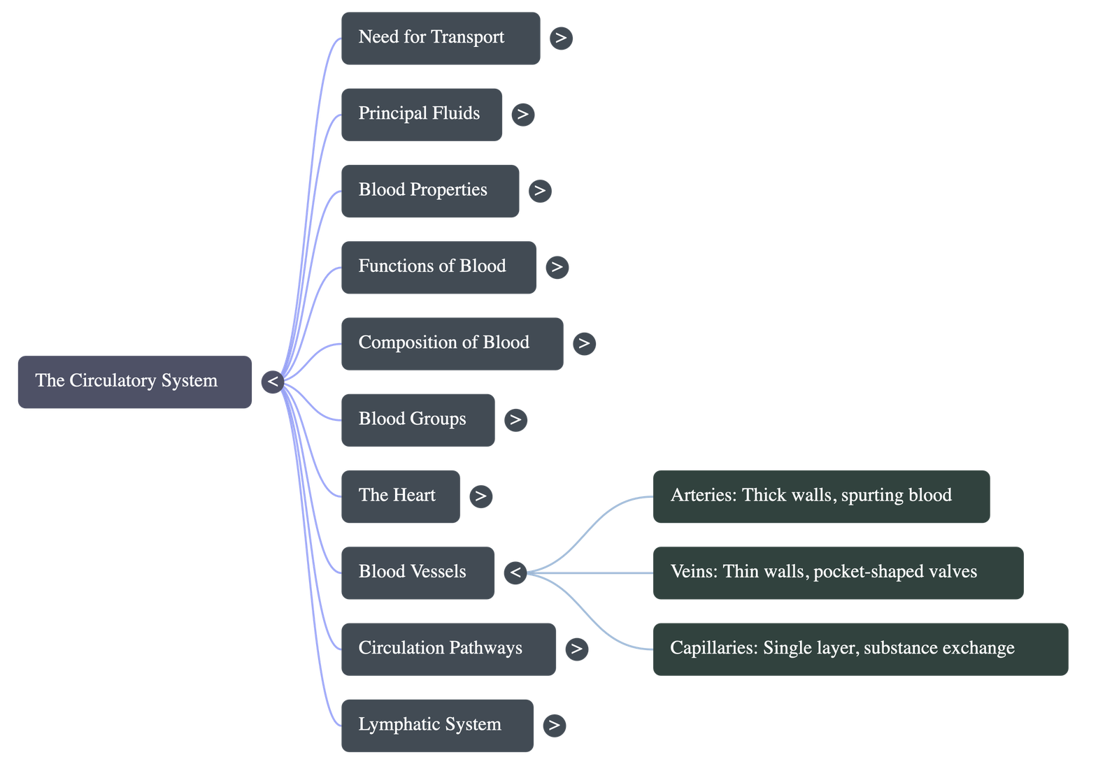
8.8.7 The Blood Vessels
- Arteries: Carry blood away from the heart. Thick muscular walls, narrow lumen, flow in spurts under high pressure, no valves.
- Veins: Carry blood to the heart. Thin muscular walls, wider lumen, smooth flow under low pressure, contain pocket-shaped valves to prevent backflow.
- Capillaries: Microscopic tubes with a single layer of squamous epithelial cells. Allow diffusion of gases, nutrients, and diapedesis of WBCs. Connect arterioles to venules.
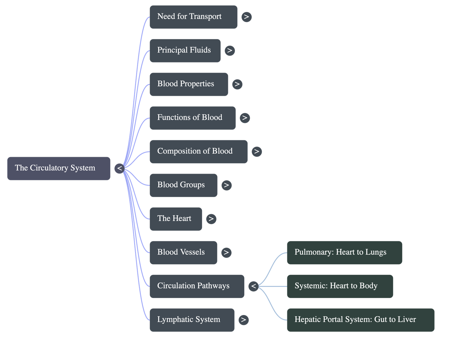
8.9 The Two Blood Circulations
- Humans have a Double Circulation system: blood flows twice through the heart in one full circuit.
- Pulmonary Circulation: Right ventricle → Lungs (oxygenation) → Left atrium.
- Systemic Circulation: Left ventricle → Entire Body tissues → Right atrium.
8.9.1 Hepatic Portal System
- Instead of going straight to the vena cava, blood from the stomach and intestines enters the liver via the Hepatic Portal Vein.
- Utility: Allows the liver to perform glycogenesis (storing excess sugar as glycogen), deamination (breaking down amino acids), and detoxification of poisons before blood reaches the heart.
8.9.2 The Pulse and Blood Pressure
- Pulse: Alternate expansion and elastic recoil of the artery wall during ventricular systole. Usually felt on the radial artery at the wrist.
- Blood Pressure: Pressure exerted by flowing blood on artery walls. Normal limit is 100-140 mm Hg (Systolic) / 60-80 mm Hg (Diastolic). Measured using a sphygmomanometer.
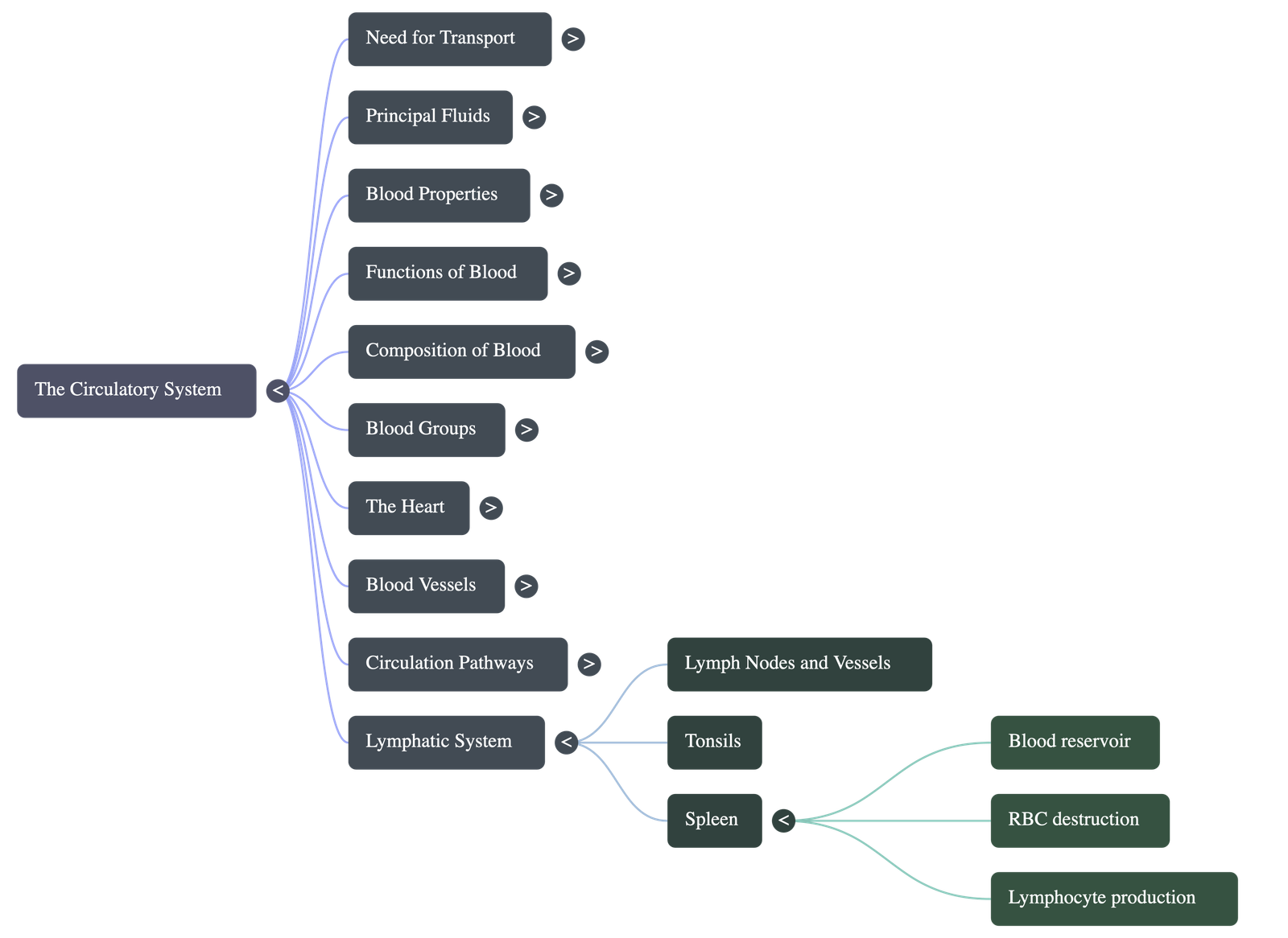
8.9.3 Tissue Fluid and Lymph
- Plasma and WBCs leak from capillaries to form Tissue fluid, exchanging substances with cells.
- Reabsorbed fluid enters lymphatic channels and becomes Lymph (contains plasma and lymphocytes, but no RBCs/platelets).
- Functions: Supplies nutrition to unreachable parts, drains excess tissue fluid, absorbs fats from intestinal villi (lacteals), and defends the body against infections.
8.9.4 The Spleen
- A large lymphatic organ behind the stomach.
- Functions: Acts as a blood reservoir (releases blood during hemorrhage or stress), produces lymphocytes, destroys worn-out RBCs, and produces RBCs in an embryo.
Do checkout all questions & answers of this chapter.
Quick Navigation:
| | | |
1 / 1
Quick Navigation:
| | | |
Quick Navigation:
| | | |