Quick Navigation:
| | | |
Sense Organs
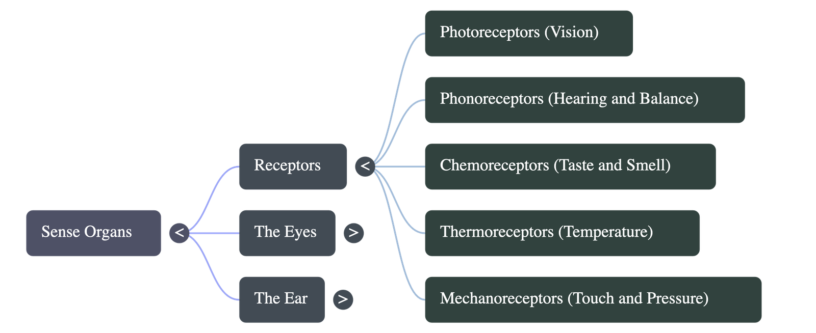
1. Introduction to Receptors
- Sense organs keep us aware of external and internal environments through specialized cells called receptors.
- The human body has five main types of receptors:
- Photoreceptors: Located in the retina of the eyes, sensitive to light (responsible for vision).
- Phonoreceptors: Located in the inner ear, sensitive to sound and balance.
- Chemoreceptors: Located in the nose and tongue, sensitive to chemicals (smell and taste).
- Thermoreceptors: Located in the skin, sensitive to relative temperature changes.
- Mechanoreceptors: Located in the skin, sensitive to mechanical stimuli like touch, pressure, and vibrations.
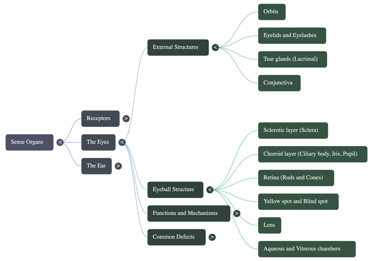
2. The Eyes: Protective Structures
- Orbits: The eyes are situated in deep bony sockets called orbits and can be rotated by six muscles.
- Eyelids and Eyebrows: Eyelids act as shutters to block out intense light, while both structures prevent rain drops and larger dust particles from entering the eye.
- Tear Glands (Lacrimal Glands): Located in the upper sideward part of the orbit, they continuously secrete fluid over the front surface. Tears lubricate the eye, wash away dust particles, communicate emotions, and contain a germ-killing enzyme called lysozyme.
- Conjunctiva: A thin, transparent membrane covering the entire front portion of the eye, reducing to a single layer of epithelium over the cornea.
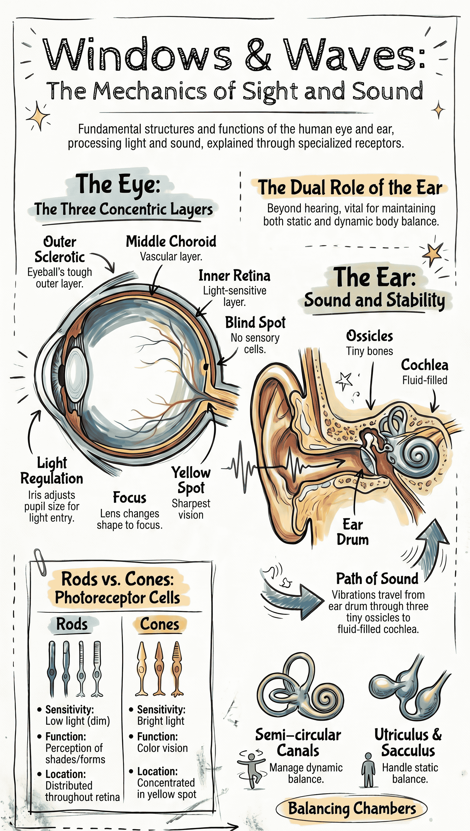
3. Structure of the Eyeball
- Sclerotic Layer (Sclera): The outermost tough, white fibrous layer that maintains eye shape. It bulges out in the front to form the transparent cornea.
- Choroid Layer: The middle layer, richly supplied with blood vessels for nourishment. It contains a dark pigment (melanin) that prevents light reflection inside the eye. In the front, it expands into the ciliary body and the iris (the colored part of the eye). The opening in the center of the iris is called the pupil.
- Retina: The innermost layer containing light-sensitive sensory cells:
- Rods: Sensitive to dim light, contain rhodopsin pigment, and do not perceive color.
- Cones: Sensitive to bright light, responsible for color vision, and contain iodopsin pigment.
4. Important Spots on the Retina
- Yellow Spot (Macula Lutea): Located at the center of the horizontal axis of the eyeball. It contains a high concentration of cones, making it the region of brightest and sharpest vision.
- Blind Spot: Located lateral to the yellow spot on the nasal side. It is the point where the optic nerve exits the eyeball; it lacks sensory cells entirely, making it an area of no vision.
5. The Lens and Eye Chambers
- Lens: A transparent, flexible, biconvex crystalline structure held in place behind the pupil by suspensory ligaments attached to the ciliary body.
- Aqueous Chamber: The space between the lens and cornea, filled with watery aqueous humour, which keeps the lens moist, protects from physical shock, and refracts light.
- Vitreous Chamber: The larger space behind the lens, filled with jelly-like vitreous humour, which helps maintain the eyeball's shape and protects the retina.
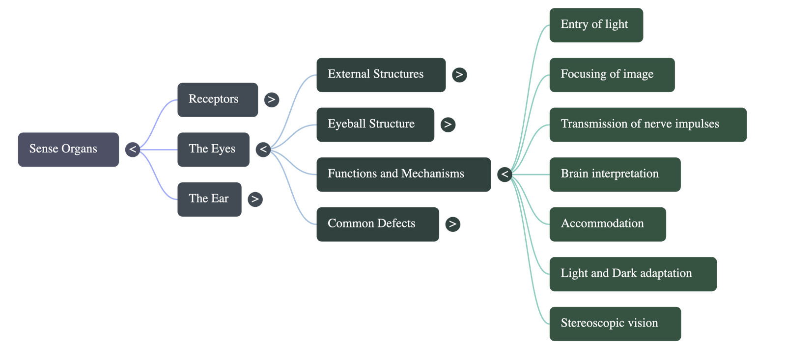
6. Vision Mechanism and Adaptation
- How We See: Light enters through transparent structures and is converged by the cornea and lens to form a real, inverted image on the retina. Chemical changes in rods and cones generate nerve impulses sent to the brain, which interprets the image upright.
- Accommodation: The ability to focus on objects at varying distances. Ciliary muscles alter the curvature and thickness of the elastic lens—flattening for distant objects and rounding out for near objects.
- Light and Dark Adaptation: Moving from bright to dim light (dark adaptation) causes rhodopsin to regenerate and the pupil to dilate. Moving from dim to bright light (light adaptation) bleaches rhodopsin and constricts the pupil to reduce light intake.
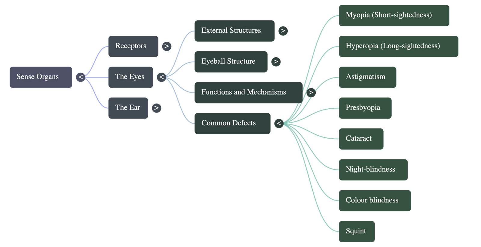
7. Common Defects of the Eye
- Myopia (Short-sightedness): Near objects are clear, distant ones blur. Image forms in front of the retina. Corrected with a concave lens.
- Hyperopia (Long-sightedness): Distant objects are clear, near ones blur. Image forms behind the retina. Corrected with a convex lens.
- Astigmatism: Blurred vision due to uneven curvature of the cornea, corrected using cylindrical lenses.
- Presbyopia: Age-related loss of lens flexibility causing far-sightedness, corrected with a convex lens.
- Cataract: The lens turns opaque leading to blindness. Corrected by surgical removal and use of high-power spectacles or a plastic lens implant.
- Night-blindness: Poor vision in dim light due to Vitamin A deficiency, which halts the synthesis of rhodopsin.
- Colour Blindness: A genetic defect causing an inability to discriminate certain colors, like red and green.
- Squint: Eyes somewhat converge ("cross eye") or diverge ("wide eye"), leading to double vision. Corrected via surgery and exercises.
- Corneal Opacities: The cornea turns white and non-functional. Corrected by replacing the defective cornea with a healthy donated one.
8. Additional Visual Phenomena
- Stereoscopic (Binocular) Vision: Allows perception of three-dimensional depth and distance due to the overlapping images formed simultaneously by both eyes.
- After-images: The sensation of an image persists on the retina for about 1/10th of a second after viewing a bright object. This biological principle enables the illusion of continuous movement in motion pictures and TV.
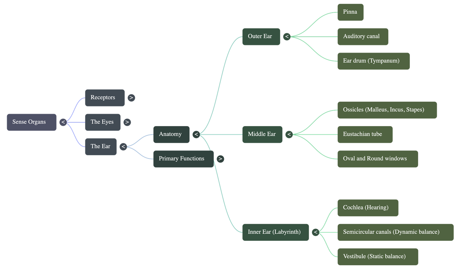
9. The Ear: Structure
- Outer Ear: Consists of the projecting flap called the pinna (auricle) and the auditory canal leading down to the ear drum (tympanum).
- Middle Ear: A cavity containing three ear ossicles—the malleus (hammer), incus (anvil), and stapes (stirrup). It is connected to the throat via the Eustachian tube to equalize air pressure on both sides of the eardrum.
- Inner Ear (Membranous Labyrinth): Composed of three main parts:
- Cochlea: A spiral snail-like tube housing the Organ of Corti, which contains sensory cells for hearing.
- Semicircular Canals: Three mutually perpendicular tubes housing sensory cells for dynamic balance.
- Vestibule: A short stem part consisting of the utriculus and sacculus containing sensory patches for static balance.
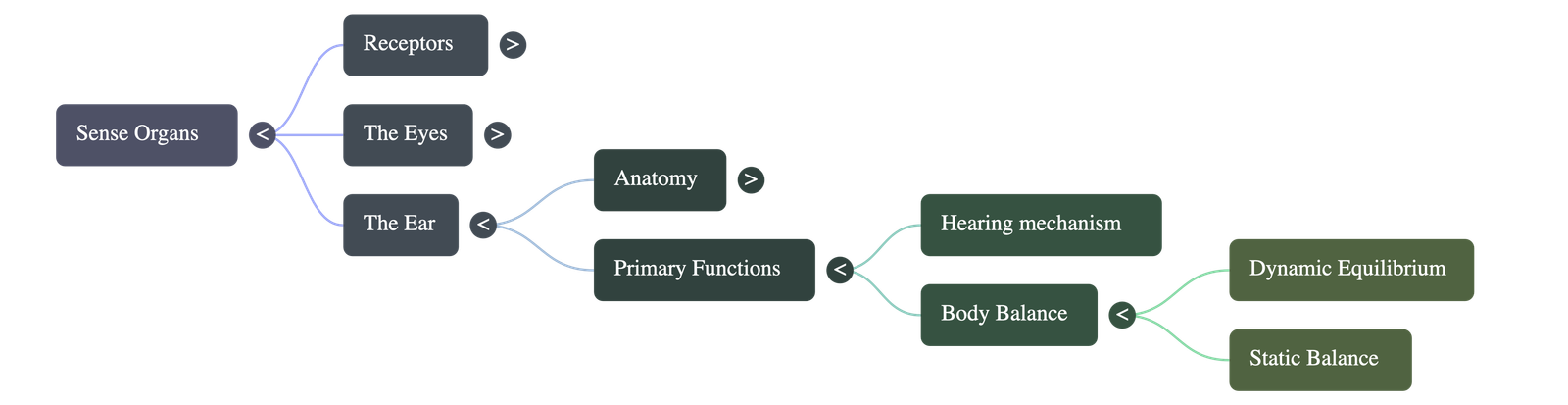
10. Functions of the Ear
- Hearing: The pinna collects sound waves that travel down the auditory canal, vibrating the eardrum. The ear ossicles strongly amplify these vibrations and transmit them to the oval window. This vibrates fluid in the cochlea, stimulating hair-like sensory cells in the Organ of Corti, which send nerve impulses to the brain via the auditory nerve.
- Balancing: The inner ear is responsible for two types of body balance:
- Dynamic Equilibrium: Fluid shifts within the semicircular canals as the head turns, pushing against sensory hair cells that send balance correction impulses when the body is in motion.
- Static Equilibrium: Sensory patches in the utriculus and sacculus perceive and register spatial orientation relative to gravity when stationary.
Do checkout all questions & answers of this chapter.
Quick Navigation:
| | | |
1 / 1
Quick Navigation:
| | | |