Quick Navigation:
| | | |
Structure of Chromosomes, Cell Cycle and Cell Division
2.1 What are Chromosomes?
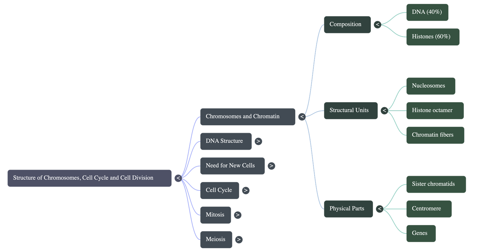
- Chromosomes are highly coiled, ribbon-like structures that appear in the nucleus only during cell division.
- When a cell is not dividing, the genetic material exists as an extremely long, thin, and darkly stained network of fibres called chromatin.
- As the cell prepares to divide, these chromatin fibres condense and coil tightly to form distinguishable chromosomes.
2.2 Discovery of Chromosomes
- First studied by German scientist Walther Flemming in 1882.
- He observed them in the rapidly dividing cells of salamander larvae.
- Flemming named the process of cell division mitosis (which literally means "thread" in Greek) because the chromosomes looked like dividing threads.
2.3 Chromatin
Chromatin fibres are composed of two main substances:
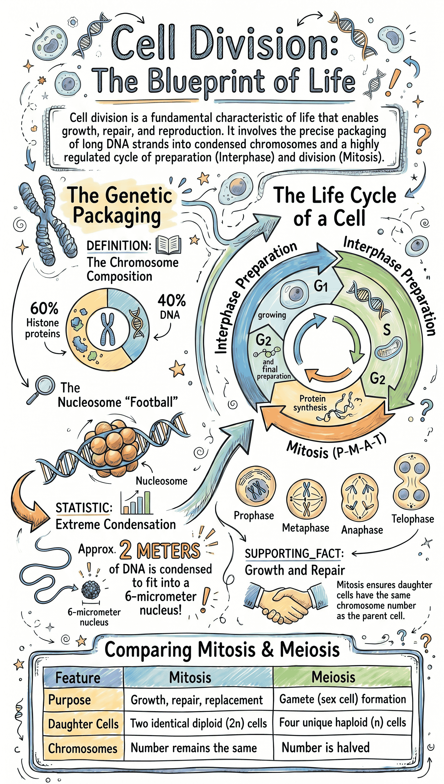
- DNA (Deoxyribonucleic Acid): Makes up about 40% of the chromatin.
- Histones: A specific type of protein making up about 60% of the chromatin.
(i) Molecular Structure of DNA
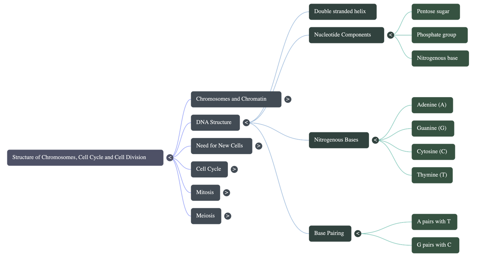
- The double-helix structure of DNA was proposed by Watson and Crick in 1953, based on studies by Rosalind Franklin.
- DNA is a macromolecule composed of two complementary strands winding around each other in a double helix.
- Each strand is made of repeating units called nucleotides.
- A nucleotide has three components: a phosphate group, a pentose sugar, and a nitrogenous base.
- The "rungs" of the DNA ladder are made of four nitrogenous bases: Adenine (A), Guanine (G), Cytosine (C), and Thymine (T).
- Pairing Rule: Adenine (A) always pairs with Thymine (T) with two hydrogen bonds. Guanine (G) always pairs with Cytosine (C) with three hydrogen bonds.
(ii) Histone Proteins
- Histones are proteins that help in coiling and packaging the exceptionally long DNA molecules so they can fit inside the tiny cell nucleus.
- Nucleosome: A structural unit formed when a DNA strand winds around a core of eight histone proteins (histone octamer).
2.4 Structure of Chromosomes
- During cell division, each visible chromosome consists of two identical halves called sister chromatids.
- These two chromatids are joined together at a small, constricted point called the centromere.
- The centromere is essential during cell division because it is the point where spindle fibres attach to pull the chromatids apart.
2.5 What are Genes?
- Genes are specific sequences of nucleotides situated on a chromosome.
- They encode instructions to produce particular proteins that express physical traits in the body.
- They are the fundamental units of heredity passed down from parents to offspring, determining specific inherited characteristics.
2.6 Need for New Cells
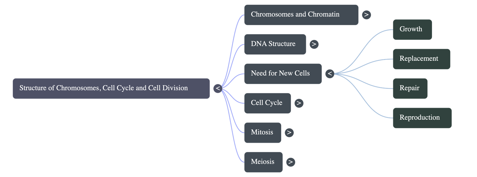
- Growth: Most organisms start as a single cell (zygote) which divides repeatedly to form tissues, organs, and ultimately the entire body.
- Replacement: Normal wear and tear destroys cells (e.g., millions of red blood cells die every second and are replaced by new ones from the bone marrow).
- Repair: In case of accidental injuries like cuts or bone fractures, cell division bridges the gap and joins the broken tissues back together.
- Reproduction: Special cell divisions produce sex cells (sperm and eggs) in higher animals, while simple organisms (like amoeba) directly divide into two to reproduce.
2.7 Cell Cycle - "Divide, grow and redivide"
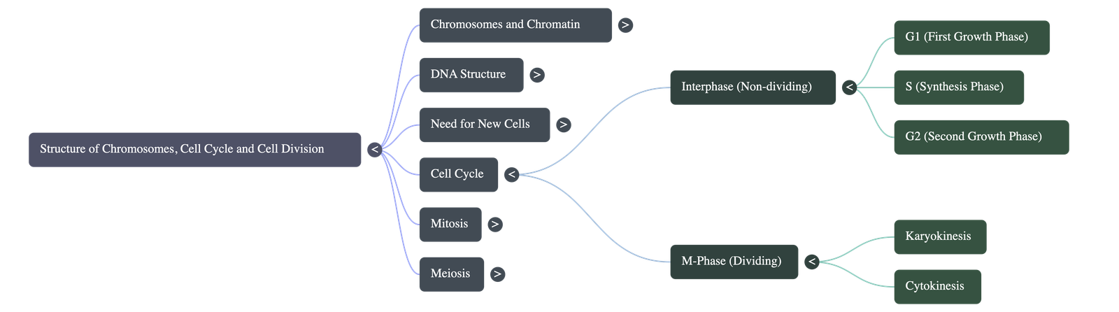
The cell cycle is an organized, predictable series of events leading to the duplication of a cell's DNA and its division into two daughter cells. It consists of two main phases:
2.7.1 Interphase
Previously misidentified as a "resting phase," Interphase is actually a highly active preparatory phase where the cell grows and synthesizes DNA. It is divided into three stages:
- First Growth Phase (G1): The cell grows in size, cytoplasm volume increases, and RNA/proteins are synthesized. Organelles like mitochondria and chloroplasts divide.
- Synthesis Phase (S): More DNA is synthesized, and the existing chromosomes duplicate to form sister chromatids (though they remain attached).
- Second Growth Phase (G2): A shorter phase where the synthesis of RNA and proteins continues, finalizing preparations for the actual division phase.
2.7.2 Formation of New DNA
- During the S-phase, the DNA double helix unzips at one end.
- New complementary strands form against each of the old, open strands, resulting in two identical DNA double helices.
2.7.3 Can the cell cycle go on endlessly?
- No, it has regulatory mechanisms.
- Brain and nerve cells stop dividing permanently after embryo formation.
- Skin and red blood cells are continuously replaced.
- If cell cycles become uncontrolled and non-stop, they may lead to the formation of tumors.
2.8 Cell Division
Cell division occurs in two major ways: Mitosis and Meiosis.
2.8.1 Mitosis
Mitosis is the division of one parent cell into two identical daughter cells, maintaining the exact same chromosome number. It occurs in two major steps:
Step A: Karyokinesis (Division of the Nucleus)
Occurs in four continuous phases:
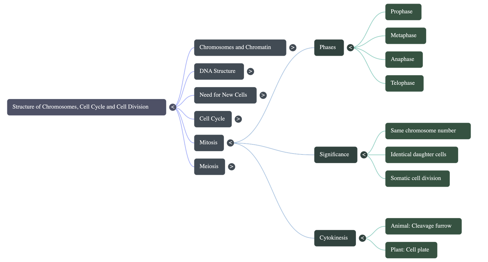
- Prophase: Chromosomes condense and become distinct. Each chromosome is already duplicated as paired chromatids attached at a centromere. Centrosomes (in animal cells) split and move to opposite poles, forming spindle fibres. The nuclear membrane and nucleolus disappear.
- Metaphase: Duplicated chromosomes align themselves along the center (equator) of the cell. Spindle fibres attach to the centromere of each chromosome.
- Anaphase: The centromeres divide. Sister chromatids separate and are pulled toward opposite poles by the contraction of spindle fibres. A furrow begins to appear in animal cells.
- Telophase: Chromatids arrive at the opposite poles and thin out to revert to chromatin threads. Spindle fibres disappear, and the nuclear membrane and nucleoli reappear.
- At the end of telophase, the cytoplasm is split into two.
2.8.2 Differences in Mitosis in Animal and Plant Cells
- Asters: Asters (star-like radiating microtubules from centrosomes) are formed in animal cells but not in plant cells.
- Cytokinesis Method: Animal cells divide cytoplasm by furrowing (membrane pinching inward). Plant cells divide by laying down a cell plate at the center, which grows outward to reach the cell walls.
- Location: Occurs in most tissues in animals; but in plants, it occurs mainly at meristematic tissues (growing tips and sides).
2.8.3 Significance of Mitosis
- Contributes to the overall growth and increased body size.
- Helps in the repair of damaged tissues.
- Replaces old and dead cells.
- Plays a role in asexual reproduction in unicellular organisms.
- Ensures the chromosome number remains identical in daughter cells.
2.9 Meiosis (Reduction Division)
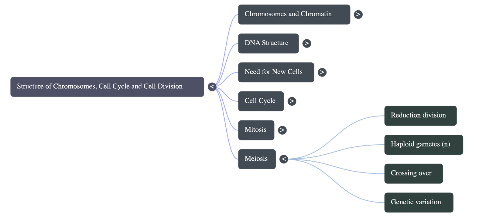
- Meiosis is a specific kind of cell division that produces sex cells or gametes (sperms and ova in humans; pollen and ovules in plants).
- Reduction: The most significant aspect of meiosis is that it halves the number of chromosomes (from a diploid "2n" state to a haploid "n" state).
- When male and female gametes fuse during fertilization, the normal diploid number of chromosomes is restored.
- Meiosis undergoes two divisions: First division separates the homologous pairs (reduction), and the second division separates the chromatids.
2.9.1 Significance of Meiosis
- Halves Chromosome Number: Ensures that upon fertilization, the offspring retains the normal chromosome number of the species rather than doubling it in every generation.
- Mixing of Genes (Genetic Recombination): Occurs via two main processes:
- Maternal and paternal chromosomes are randomly mixed during separation.
- Crossing Over: During division, non-sister chromatids of a paired homologous chromosome exchange genetic material at attachment points known as chiasmata.
- This continuous mixing of genes is the root cause of the vast variations seen among children of the same parents and within a species.
Do checkout all questions & answers of this chapter.
Quick Navigation:
| | | |
1 / 1
Quick Navigation:
| | | |